
Congenital Foot Deformities in Children
Specialized pediatric care for clubfoot, accessory navicular syndrome, and other congenital foot conditions.
What You Need To Know
Understanding congenital foot deformities and how early treatment supports healthy development.
Pediatric Congenital Foot Deformities
Congenital foot deformities are conditions present at birth that affect the shape, structure, or function of a child’s foot. Two of the most common conditions we treat are clubfoot and accessory navicular syndrome. Early evaluation allows us to take advantage of the flexibility of a child’s developing bones, tendons, and joints.
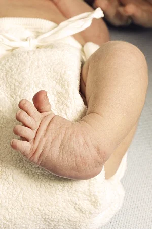
Clubfoot is one of the most common congenital deformities, occurring in about 1 out of every 1000 births. It affects males more commonly than females. The foot shape at birth is pointed inward and downward, giving the foot a club appearance. The tendons in the baby’s foot are shorter, so the foot is out of position or twisted. Signs include a foot twisted inward and downward, a foot that appears upside down, a shorter foot or leg, and underdeveloped calf muscles. Usually there is no pain.
The accessory navicular is an extra bone or cartilage located on the inner side of the foot just above the arch, incorporated within the posterior tibial tendon. Many people are unaware of the condition if it causes no problems. However, some develop a painful condition known as accessory navicular syndrome when the bone or tendon is aggravated by trauma, chronic irritation from footwear, or overuse. Adolescence is a common time for symptoms to first appear.
To diagnose these conditions, your doctor will ask about symptoms and examine the foot, evaluating structure, muscle strength, joint motion, and gait. X-rays are usually ordered to confirm the diagnosis. If your child shows signs of a congenital foot deformity, we encourage you to schedule an evaluation so treatment can begin at the most effective time.
Treatment Options and the Ponseti Method
Treatment for clubfoot should begin in the first few weeks after birth when the baby’s joints, bones, and tendons are flexible. Stretching and casting is the most common treatment and is called the Ponseti method. The doctor moves the baby’s foot to the correct position and applies a cast. The cast is changed and the foot repositioned about once a week for several months. The Achilles tendon may need to be lengthened, and stretching exercises, braces, and special shoes help maintain the corrected position.
Dr. Brianna David, who has taken over her father Dr. Michael David’s clubfoot practice, utilizes the Ponseti Method of serial casting. Treatment should begin within 1 to 2 weeks after birth. The foot is gently manipulated and plaster casts are applied and changed weekly for 3 to 6 weeks in the office without anesthesia. Correction can be achieved in over 90 percent of cases without extensive surgery. Occasionally a release of the heel cord is needed, performed in the office through a tiny incision.
Relapses are uncommon and are usually corrected with 1 to 3 more casts. Special shoes attached to a bar are used 20 to 22 hours per day for 3 months, followed by nighttime use for 2 to 4 years.
For accessory navicular syndrome, nonsurgical care includes immobilization in a cast or walking boot, ice, NSAIDs, physical therapy, and orthotic devices to support the arch. If nonsurgical treatment fails, surgery may involve removing the accessory bone, reshaping the area, and repairing the posterior tibial tendon. With early, appropriate care, most children go on to walk, run, and play comfortably.
Pediatric Foot Care Near You
Find a SoCal Foot Care location that offers pediatric congenital foot deformity treatment.









If you’re concerned about your child’s foot shape or development, let’s take a closer look together.
Early evaluation makes a difference. We’re here to help your child thrive.